Order Code
142
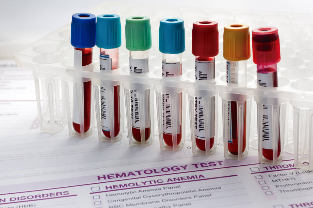
Preferred Specimen
Collect 2 mL of serum. Allow the SST tube to clot upright for at least 30 minutes, then centrifuge within 2 hours of collection. Store refrigerated.
ContainerType
Serum separator tube
Alternate Specimen Requirements
Collect 2 mL of serum using a plain red-top tube. Allow the specimen to clot upright for a minimum of 60 minutes. Then centrifuge, transfer the serum into a plastic transport tube within 2 hours, and clearly label it as “serum from red-top tube.” Store refrigerated.
Minimum Volume
2 mL
Transport Temperature
Refrigerated
Expected Turnaround Time
1 day room temperature; 1 week refrigerated
Specimen Stability
Fresh urine:1 day refrigerated BD urine preservative tube:3 days room temperature
Methodology
Roche COBAS automated methodology or other
automated chemistry method.
Overview
The Basic Metabolic Panel (BMP) consists of seven or eight biochemical assays that assess electrolyte balance, renal function, and glucose concentration. When interpreted in concert, abnormal results may provide insights into potential disease processes requiring further investigation (AACC, 2020).
A BMP typically includes:
- Sodium (Na⁺) — primarily extracellular; plays a central role in regulating water distribution
- Potassium (K⁺) — primarily intracellular; small plasma changes can significantly affect cardiac function
- Chloride (Cl⁻) — moves with sodium across cell membranes to support electrical and osmotic equilibrium
- Bicarbonate (HCO₃⁻) — helps maintain acid–base balance through renal mechanisms
- BUN (Blood Urea Nitrogen) — a byproduct of protein metabolism, filtered by the kidneys; used as a marker of renal function
- Creatinine (Cr) — derived from muscle metabolism; indicates renal filtration capacity
- Calcium (Ca²⁺) — mostly stored in bone; circulating levels are tightly controlled hormonally
- Glucose (Glc) — the body’s principal fuel source, regulated by diet, hormones, and metabolism
Some laboratories may choose not to report calcium depending on their BMP panel configuration.
Clinical Significance
- Used in routine health screening
- Detects disturbances in fluid or electrolyte balance
- Helps assess acid–base imbalances
- Aids in diagnoses when symptoms such as edema, nausea, weakness, confusion, or arrhythmia are present
- Monitors acute or chronic conditions (e.g., hypertension, heart failure, liver or kidney disease, diabetes)
- Evaluates effects of medications that may alter electrolyte levels (e.g., diuretics)
Additional Information
Although sodium, potassium, chloride, and bicarbonate are often measured as a group (electrolyte panel), each can be ordered individually when clinically indicated. Other essential electrolytes—such as calcium, magnesium, and phosphate—may also be requested separately (AACC, 2019).
Interpretative Information
Sodium:
Hypernatremia (>150 mEq/L)
- Common causes:
- Dehydration (eg, diuretics, vomiting or diarrhea, increased free water loss from fever, sweating, or burns)
- Drug side effect (eg, anabolic steroids, corticosteroids, laxatives, cough medicines, and oral contraceptives)
- Less obvious causes:
- Cushing syndrome
- Central or nephrogenic diabetes insipidus with insufficient free water
- Hyperaldosteronism
- Excessive salt intake
Hyponatremia (<125 mEq/L)
- Common causes:
- Hypervolemic
- Dilution due to water retention (eg, decompensated heart failure, advanced liver cirrhosis, renal failure)
- Euvolemic
- Hypothyroidism
- Syndrome of inappropriate secretion of antidiuretic hormone (SIADH)
- Glucocorticoid deficiency
- Beer potomania
- Psychogenic polydipsia
- Hypovolemic
- Diarrhea or vomiting
- Diuretic use
- Mineralocorticoid deficiency
- Third spacing (ileus, pancreatitis)
- Hypervolemic
- Pseudohyponatremia may occur in the presence of very high glucoses (eg, diabetic ketoacidosis or nonketotic hyperosmolar coma)
- Sodium is artificially decreased by 1.3-1.6 mEq/L for each 100 mg/dL increase in glucose. Apparent mild hyponatremia with very high glucose may actually mean hypernatremia.
Potassium:
Hyperkalemia (>6.5 mEq/L)
- Excessive intake or administration
- Medications (eg, K+ salts of some drugs, ACE inhibitors, NSAIDs, Aldactone, high-dose trimethoprim-sulfamethoxazole therapy)
- Mobilization of potassium from cells (eg, trauma, malignant hyperthermia)
- Inadequate renal excretion (renal insufficiency)
- Addison disease
- Acidosis (eg, diabetic ketoacidosis or lactic acidosis)
- Lack of insulin
- Renal tubular disorders
- Artifact causing hyperkalemia includes hemolysis from collection, storage, or processing
Hypokalemia (<2.5 mEq/L)
- Drug-induced (eg, diuretics, bronchodilators, mineralocorticoids, high-dose glucocorticoids, high-dose penicillin, aminoglycosides, cisplatin, foscarnet, amphotericin B)
- Found in 80% to 90% of hypertensive patients with primary aldosteronism
- Parenteral deprivation of K+ (eg, parenteral therapy without adequate K+ replacement)
- GI loss (vomiting, diarrhea, bypass, enteric fistulas, malabsorption)
- Malnutrition (including alcoholism)
- Alkalosis
- Renal tubular disorders
Chloride:
Hyperchloridemia (>115 mEq/L)
- Hyperchloremic (nonanion gap) metabolic acidosis
- Excessive infusion of hyperchloremic (normal) saline
- Diarrhea/GI losses
- Renal tubular acidosis
- Pancreatic fistula
- Enterovesical fistula
- Mineralocorticoid deficiency
Hypochloridemia (<80 mEq/L)
- Vomiting or gastric suction
- Overhydration
- Syndrome of inappropriate secretion of ADH
- Chronic or compensated respiratory acidosis
- Burns
- Diuretic therapy
Bicarbonate: Along with anion gap (see Pearls), HCO3– is used as a preliminary screen for abnormalities in acid-base balance:
Elevated HCO3– (>30 mEq/L)
- Metabolic alkalosis, usually acute and often accompanied by hypokalemia
- Compensated respiratory acidosis
- Increased cortisol or aldosterone causing metabolic alkalosis from urinary losses of hydrogen and potassium ions
Decreased HCO3– (<10 mEq/L)
- Metabolic acidosis
- Compensated respiratory alkalosis
- Hyperchloremic (nonanion gap) metabolic acidosis − decreased HCO3– with high chloride and normal anion gap (eg, diarrhea and renal tubular acidosis)
- Drug-induced (eg, methicillin, nitrofurantoin, tetracycline, thiazide diuretics)
BUN (blood urea nitrogen):
Elevated BUN (>100 mg/dL)
- Kidney disease
- Urinary tract obstruction (stone, stricture, external compression)
- Decreased blood flow to the kidneys (dehydration, heart failure, shock, severe burns)
- Gastrointestinal bleeding (upper GI such as stomach, esophageal)
- Increased protein breakdown (sepsis, fevers)
- High protein diets
- Increasing age (in particular, over age 60)
- Drug-induced (eg, aminoglycosides, cephalosporins, diuretics, steroids)
Decreased BUN (<7 mg/dL)
- Uncommon and not usually a cause for concern
- Malnutrition (eg, cancer, advanced liver disease, alcoholism)
- Overhydration
- Drug-induced (eg, chloramphenicol, streptomycin)
Creatinine:
Elevated (>8 mg/dL)
- Prerenal causes of reduced blood flow to the kidney (dehydration, heart failure, shock, renal artery stenosis)
- Damage to renal vasculature (diabetes, hypertension, glomerulonephritis due to infection or autoimmune diseases)
- Bacterial infection of kidneys (pyelonephritis)
- Damage to renal collecting tubules (acute tubular necrosis due to drugs, shock, or toxins)
- Postrenal obstruction (kidney stone, prostate hypertrophy)
Decreased:
- Uncommon and are not usually a cause for concern
- May be seen in decreased muscle mass
Glucose:
Hyperglycemia (>99 mg/dL)
- Diabetes mellitus, I or II
- Acute physiologic stress (trauma, myocardial infarction, infection)
- Endocrinopathies (eg, acromegaly, Cushing disease or syndrome, hyperthyroidism)
Hypoglycemia (<65 mg/dL)
- Hepatic dysfunction (eg, cirrhosis)
- Severe heart failure
- Sepsis
- Endocrinopathies (eg, hypothyroidism, hypopituitarism)
- Tumors (insulinomas)
- Accidental or deliberate misuse of hypoglycemics (insulin or oral glucose-lowering medications)
Calcium:
Hypercalcemia (>10.3 mg/dL)
- Hyperparathyroidism
- Cancer (excessive secretion of parathyroid hormone-related protein, bony metastasis)
- Disease (eg, sarcoidosis, tuberculosis)
- Prolonged immobilization
- Drug-induced (eg, thiazide diuretics, lithium, tamoxifen, excessive vitamin D intake)
Hypocalcemia (<8.5 mg/dL)
- Hypoalbuminemia (most common; results in a decrease in bound calcium, ionized calcium levels remains normal)
- Hypoparathyroidism
- Renal failure
- Deficiency of dietary calcium, magnesium, or vitamin D
- Increased levels of phosphorus
- Acute pancreatitis
Limitations
Potassium:
- Falsely elevated reading due to blood cell hemolysis from poor collection technique causing the lysis of cells and subsequent release of high concentrations of intracellular potassium.
- Falsely elevated reading due to prolonged contact of serum with cells if a specimen has sat unattended for several hours.
Bicarbonate:
- Falsely low reading if specimen is exposed to air causing loss of carbon dioxide
- Falsely low reading from inadequate filling of red top evacuated collection tubes
- Falsely low reading (as much as 10 mmol/L) if specimen is excessively diluted with heparin (use only enough heparin sufficient to fill the dead space of the syringe)
Creatinine:
- Falsely low reading when high concentrations of NAPQI (an acetaminophen metabolite) or N-acetylcysteine (NAC) are present
- Normal serum creatinine results do not rule out renal dysfunction as the serum creatinine is not sensitive to early renal damage.
References
American Association for Clinical Chemistry (AACC). Basic Metabolic Panel (BMP). AACC website. https://labtestsonline.org/tests/basic-metabolic-panel-bmp. Updated July 29, 2020. Accessed August 23, 2020.
American Association for Clinical Chemistry (AACC). Calcium. AACC website. https://labtestsonline.org/tests/calcium. Updated July 19. 2017. Accessed August 25, 2020.
American Association for Clinical Chemistry (AACC). Electrolytes and Anion Gap. AACC website. https://labtestsonline.org/tests/aldosterone-and-renin. Updated September 5, 2019. Accessed August 14, 2020.
Freda BJ, Davidson MB, Hall PM. Evaluation of hyponatremia: a little physiology goes a long way. Cleve Clin J Med. 2004;71(8):639-650. doi:10.3949/ccjm.71.8.63915449759
Kapsner CO, Tzamaloukas AH. Understanding serum electrolytes. How to avoid mistakes. Postgrad Med. 1991;90(8):151-161. doi:10.1080/00325481.1991.117011461749730
Lee S, Kang KP, Kang SK. Clinical usefulness of the serum anion gap. Electrolyte Blood Press. 2006;4(1):44-46. doi:10.5049/EBP.2006.4.1.4424459484
Nickson C. Anion Gap. Life in the Fastlane website: https://litfl.com/anion-gap. Update April 23, 2019. Accessed July 16, 2020.
Scott MG, Heusel JW, LeGrys VA, et al. Electrolytes and Blood Gases. In: Burtis CA, Ashwood ER, eds. Tietz Textbook of Clinical Chemistry. 3rd ed. Philadelphia, PA: WB Saunders Co; 1999:1056-92.
Diagnostic Role
The basic metabolic panel (BMP) is ordered during both routine health exams and in acute evaluations to identify electrolyte, fluid, metabolic imbalance (acidosis or alkalosis), renal function, and/or glucose abnormalities. The BMP is used to screen for conditions such as diabetes or renal disease, as well as monitor known conditions, such as hypertension (AACC 2020).
Results are evaluated for both individual lab abnormalities as well as together to identify patterns that may identify or suggest underlying pathology, such as dehydration, renal or pulmonary disease, or endocrine abnormalities. A series of BMPs may be followed over time to monitor interventions (AACC 2019). If the resulted values do not correlate with clinical findings, hemolysis or laboratory error needs to be ruled out, typically by resubmitting a new blood sample for the laboratory to analyze.
Alias
- Bmp
Test Setup Days
Monday through Friday
CPT
80048 AMA Defined Panel.