Order Code
2776
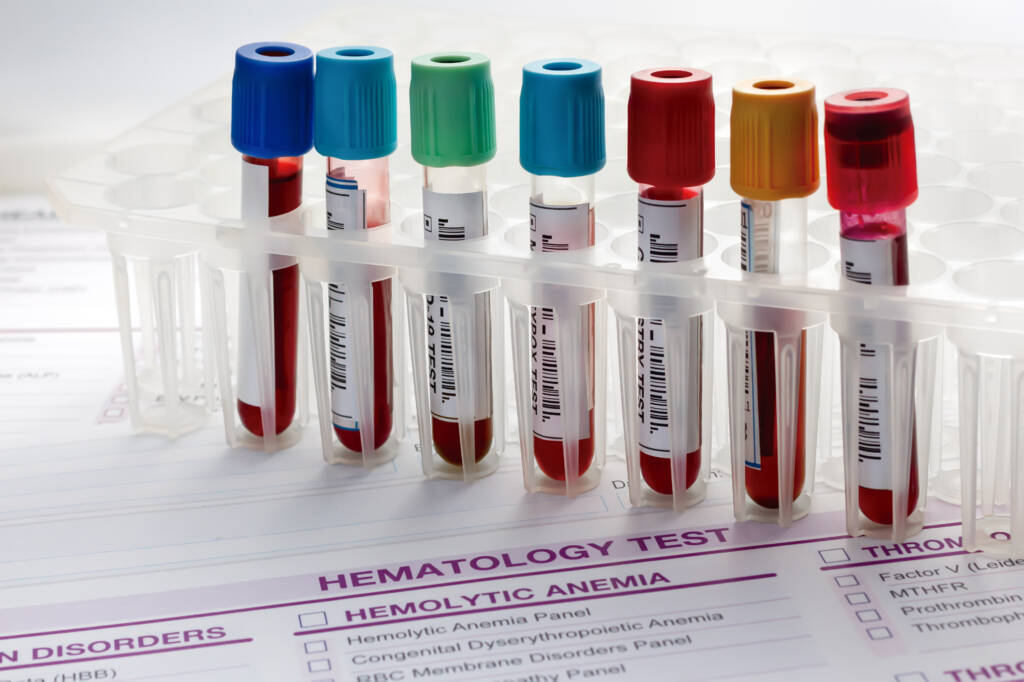
Preferred Specimen
- 2 mL serum collected in SST tube
- Allow clotting in an upright position for at least 30 minutes
- Centrifuge within 2 hours of collection
- Refrigerate after processing
- Important: Do not collect if patient is on high-dose biotin therapy (>5 mg/day) until 8 hours after last dose
ContainerType
Serum separator tube
Alternate Specimen Requirements
- 2 mL serum from a plain red top tube
- Allow clotting upright for at least 60 minutes
- Centrifuge and transfer serum to plastic transport tube within 2 hours
- Label clearly as serum from plain red top tube
- Refrigerate
- Important: Same biotin restriction applies
Minimum Volume
Adult: 0.5 mL serum
Pediatric: 0.3 mL serum (does not allow for repeat or
additional testing).
Transport Temperature
Refrigerated
Expected Turnaround Time
1-2 days
Specimen Stability
5 days room temperature; 2 weeks refrigerated; 6 months frozen. Allow only one freeze/thaw cycle
Methodology
Roche COBAS Electrochemiluminescent Immunoassay (ECLIA) this method has been standardized against the 2nd international standard (NIBSC) 80/552. Note: STAT or regional laboratories may use different methodology and/or manufacturer.
Overview
Luteinizing hormone (LH) a is glycoprotein gonadotropic hormone produced by the pituitary gland. It is composed of an α and β subunit. The α subunit is identical to that of follicle stimulating hormone (FSH), thyroid stimulating hormone (TSH), and human chorionic gonadotropin (hCG). Specificity resides in the β subunits. In females, LH promotes the production of progesterone and androgens in the ovary. It is also involved in ovulation. In males, LH stimulates the production of testosterone and is involved in spermatogenesis along with FSH.
Clinical Significance
- Evaluation of menstrual irregularities:
- Primary amenorrhea (absence of menses by age 16 with secondary sexual characteristics or by age 14 without secondary sexual characteristics)
- Secondary amenorrhea (absence of menses for 6 months in a previously menstruating female)
- Oligomenorrhea (menses more than 35 days apart)
- Anovulatory bleeding
- Precocious puberty
- Evaluation of infertility
- Evaluation of male hypogonadism
Additional Information
Females:
LH is secreted by the pituitary gland along with FSH in response to pulsatile release of gonadotropin releasing hormone (GnRH) from the hypothalamus. Puberty is marked by the nocturnal increase in LH secretion caused by an increase in GnRH secretion. Both FSH and LH secretion vary over the course of the menstrual cycle. During the follicular phase of the menstrual cycle (first day of menses through ovulation), FSH stimulates the growth of ovarian follicles, especially the dominant follicle, and induces production of estrogen. Once estrogen levels reach a certain value (usually >200 pg/mL), a surge in LH is stimulated and ovulation occurs. LH then supports the corpus luteum and production of progesterone during the luteal phase (ovulation to start of menses).
FSH and LH are under complex regulation by hypothalamic GnRH and by gonadal sex hormones (estrogen, progesterone). On the simplest level, when estrogen levels are low, FSH is released to stimulate ovarian follicle development and estrogen production. When estrogen levels are elevated, FSH release is suppressed. Estrogen has the opposite effect on LH. When estrogen levels are high, LH is released to trigger ovulation. When estrogen levels are low, LH is suppressed. LH stimulates production of progesterone; when progesterone levels rise it then has an inhibitory effect on LH.
LH measurements are used during the evaluation and diagnosis of precocious puberty. It is especially useful in distinguishing gonadotropin-dependent from gonadotropin–independent precocious puberty. Basal and GnRH-stimulated serum levels of LH are obtained. If LH levels are elevated, then gonadotropin-dependent precocious puberty is likely.
LH increases in urine after the pituitary’s LH surge that precedes ovulation by 24-36 hours. Home use test kits for qualitative urine LH are available for predicting when ovulation is likely to occur and have been reported to be effective for this use.
Males:
Again, LH is secreted by the pituitary gland along with FSH in response to pulsatile release of gonadotropin releasing hormone (GnRH) from the hypothalamus. Both FSH and LH play an important role in normal spermatogenesis. LH stimulates the Leydig cells of the testis to produce testosterone while FSH stimulates the Sertoli cells. LH secretion is controlled through negative feedback by testosterone.
Interpretative Information
During the evaluation of amenorrhea and anovulation in females and hypogonadism in males, FSH, LH, and sex steroid levels (estrogen, progesterone, and testosterone) can be placed into three categories. In general, FSH and LH are elevated in conditions in which sex hormones cannot be elaborated and are decreased in conditions of hypothalamic/pituitary dysfunction.
Females:
- Hypergonadotropic hypogonadism: Elevated FSH, LH with decreased estrogen/progesterone:
- Seen with gonadal failure: Turner’s syndrome and other chromosomal abnormalities, gonadal dysgenesis, premature ovarian failure (chemotherapy, gonadal radiation, genetic disorders, autoimmune disorders), menopause
- Hypogonadotropic hypogonadism: FSH, LH, estrogen, progesterone are decreased
- Examples include congenital GnRH deficiency; pituitary gonadotropin deficiency; pituitary adenomas; Kallman’s syndrome; hyperprolactinemia; hypothyroidism; and extreme stress, exercise, or eating disorders
- Eugonadism: FSH, LH, estrogen, and progesterone levels are all normal
- Chronic anovulation, such as polycystic ovarian disease, hyperprolactinemia, hypothyroidism, obesity. In polycystic ovarian disease, LH:FSH ratio is >2:1, but this is not used for diagnosis.
- Anatomic defects: Asherman’s syndrome, muellerian agenesis, labial agglutination
Males:
- Hypergonadotropic hypogonadism (elevated FSH, LH; low testosterone):
- Klinefelter’s syndrome; cryptorchidism; testicular failure from chemotherapy, radiation, alcohol abuse, testicular injury, and infection
- Hypogonadotropic hypogonadism (low FSH, LH; low testosterone):
- Hyperprolactinemia, Kallman’s syndrome, hypothalamic and pituitary tumors and disorders
Limitations
- In females, both FSH and LH vary over the course of the menstrual cycle; thus, interpretation of a single determination may be difficult and must be in relation to time in menstrual cycle.
- Increased LH with normal or low FSH may occur with obesity, hyperthyroidism, and in liver disease.
- Normal LH and FSH (RIA) levels can occur in hypoestrogenic patients and in patients with CNS/pituitary failure.
- The glycoprotein hormones can be heterogeneous inactive molecules which may circulate and cross-react with reagent antibodies in the radioimmunoassay to give a false value.
References
Burger HG, Hale GE, Robertson DM, et al. A review of hormonal changes during the menopausal transition: focus on findings from the melbourne women’s midlife health project. Human Reprod Update. 2007;13(6):559–565.17630397
Cleveland Clinic Foundation, Test Directory, 2013.
Fritz MA, Speroff L. Amenorrhea. Clinical Gynecologic Endocrinology and Infertility. 8th ed. Philadelphia, PA: Lippincott Williams and Wilkins;2011.
Fritz MA, Speroff L. Neuroendocrinology. Clinical Gynecologic Endocrinology and Infertility. 8th ed. Philadelphia, PA: Lippincott Williams and Wilkins;2011.
Fritz MA, Speroff L. Normal and abnormal growth and pubertal development. Clinical Gynecologic Endocrinology and Infertility. 8th ed. Philadelphia, PA: Lippincott Williams and Wilkins;2011.
Fritz MA, Speroff L. Regulation of the menstrual cycle. Clinical Gynecologic Endocrinology and Infertility. 8th ed. Philadelphia, PA: Lippincott Williams and Wilkins;2011.
Nguyen HCT. Luteinizing Hormone. Medscape. 2014.
Miller PB, Soules MR. The usefulness of a urinary LH kit for ovulation prediction during menstrual cycles of normal women. Obstet Gynecol. 1996; 87(1):13-17.8532248
Wu AHB, ed. Tietz Clinical Guide to Laboratory Tests. 4th ed. St Louis, MO: Saunders/Elsevier; 2006:694-696.
Alias
- D6706
- Lh
Test Setup Days
Monday through Friday PM shift
CPT
83002 LOINC: 10501-5
Reference Range
MALES FEMALES
AGE: 7-9 YEARS <=0.7 IU/L <=0.7 IU/L
10-12 YEARS <=3.4 IU/L <=6.8 IU/L
13-15 YEARS 0.3-5.6 IU/L <=23.0 IU/L
16-17 YEARS 1.1-9.0 IU/L <=26.4 IU/L
>=18 YEARS 1.2-8.6 IU/L
FOLLICULAR 2.4-12.6 IU/L
MID-CYCLE PEAK 14.0-95.6 IU/L
LUTEAL PHASE 1.0-11.4 IU/L
POSTMENOPAUSAL 7.7-58.5 IU/L
MALES FEMALES
TANNER I <=1.0 IU/L <=2.8 IU/L
TANNER II <=3.6 IU/L <=7.9 IU/L
TANNER III <=6.4 IU/L <=23.0 IU/L
TANNER IV/V 1.1-8.5 IU/L <=25.3 IU/L
| UNIT CODE | UNIT CODE NAME | ANALYTE | GENDER | AGE | REFERENCE RANGE | Units of Measure |
|---|---|---|---|---|---|---|
| 2776 | LH | LH | NOT SPECIFIED | 0Y | SEE BELOW | IU/L |
| 2776 | LH | LH | NOT SPECIFIED | 17Y | SEE BELOW | IU/L |
| 2776 | LH | LH | NOT SPECIFIED | 150Y | SEE BELOW | IU/L |
| 2776 | LH | LH | MALE | 0Y | 1.2-8.6 | IU/L |
| 2776 | LH | LH | MALE | 17Y | SEE BELOW | IU/L |
| 2776 | LH | LH | MALE | 150Y | 1.2-8.6 | IU/L |
| 2776 | LH | LH | FEMALE | 0Y | SEE BELOW | IU/L |
| 2776 | LH | LH | FEMALE | 17Y | SEE BELOW | IU/L |
| 2776 | LH | LH | FEMALE | 150Y | SEE BELOW | IU/L |