Order Code
9324
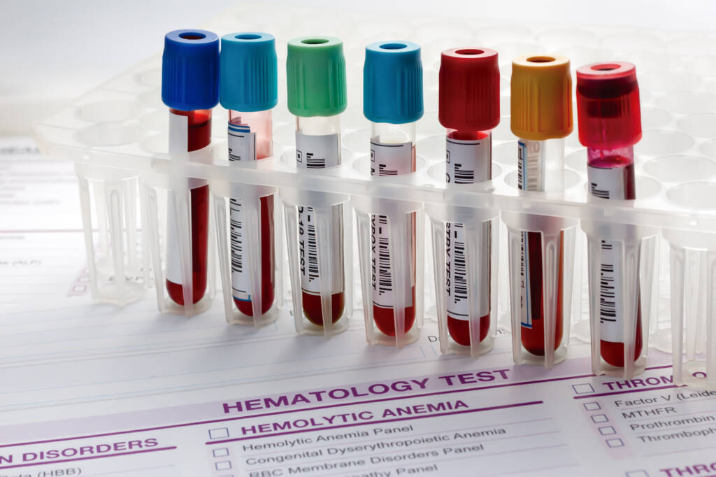
Preferred Specimen
Collect 2 mL of serum. Allow the SST (serum separator tube) to clot upright for a minimum of 30 minutes, then centrifuge within 2 hours of collection. Store refrigerated.
ContainerType
Serum separator tube
Alternate Specimen Requirements
Collect 2 mL of serum using a plain red-top tube. Permit the sample to clot upright for at least 60 minutes. Centrifuge and transfer the serum into a labeled plastic transport tube within 2 hours of collection. Clearly indicate that the specimen is from a plain red-top tube. Keep refrigerated.
Minimum Volume
2 mL serum
Transport Temperature
Refrigerated
Specimen Stability
1 day room temperature; 1 week refrigerated
Methodology
Roche COBAS automated methodology or other
automated chemistry method.
Overview
The kidney function panel comprises ten biochemical assays that evaluate fluid and electrolyte balance, renal performance, and levels of glucose and albumin. Deviations from the reference range—particularly in a patterned manner—may signal an underlying condition requiring further clinical investigation (AACC Renal Panel, 2019).
Albumin: A major plasma protein produced by the liver, representing about 60% of total serum protein.
Sodium (Na⁺): Primarily extracellular, sodium regulates water distribution and fluid balance.
Potassium (K⁺): Mainly intracellular, but even small elevations in plasma levels can significantly affect cardiac rhythm and muscle function.
Chloride (Cl⁻): Moves alongside sodium to help maintain electrical neutrality and fluid homeostasis.
Bicarbonate (HCO₃⁻): Critical for acid-base balance, regulated through renal excretion and reabsorption.
BUN (Blood Urea Nitrogen): A byproduct of protein metabolism filtered by the kidneys, used to assess renal function.
Creatinine (Cr): Derived from muscle metabolism, creatinine levels provide another marker of kidney filtration efficiency.
Calcium (Ca²⁺): Mostly stored in bone, with tightly controlled circulating levels influenced by hormonal regulation.
Phosphorus (PO₄³⁻): Predominantly intracellular; involved in energy metabolism, neuromuscular function, and bone health.
Glucose (Glc): The body’s main energy source, regulated through a balance of diet, hormones, and metabolic demands.
Clinical Significance
- Evaluation of kidney function
- Evaluation of imbalances in the body’s fluid, electrolyte and acid-base status given known kidney dysfunction
Additional Information
The electrolytes—sodium, potassium, chloride, bicarbonate, calcium, and phosphorus—can also be ordered individually for targeted diagnostic purposes. Magnesium, another essential electrolyte, is available as a separate test.
Interpretative Information
Sodium
- Hypernatremia (>150 mEq/L)
Common causes: Dehydration (diuretics, vomiting, diarrhea), excessive water loss (fever, burns), certain medications (steroids, laxatives), Cushing syndrome, diabetes insipidus, hyperaldosteronism, high salt intake.
Note: Glucose elevation can cause a falsely low sodium; expect a 1.3–1.6 mEq/L decrease per 100 mg/dL glucose. - Hyponatremia (<125 mEq/L)
Common causes:- Hypervolemic: CHF, cirrhosis, renal failure
- Euvolemic: SIADH, hypothyroidism, glucocorticoid deficiency
- Hypovolemic: GI losses, diuretics, adrenal insufficiency
Pseudohyponatremia may occur with high serum lipids or proteins, or significantly elevated glucose.
Potassium
- Hyperkalemia (>6.5 mEq/L)
Causes include increased intake, renal impairment, medications (ACE inhibitors, NSAIDs, Aldactone), cell lysis (trauma), and acidosis. Hemolysis during collection can cause artifactual elevation. - Hypokalemia (<2.5 mEq/L)
Commonly drug-induced (diuretics, corticosteroids, etc.), also from GI losses, malnutrition, alkalosis, or renal tubular defects. Seen in most hypertensive patients with primary aldosteronism.
Chloride
- Hyperchloremia (>115 mEq/L)
Associated with metabolic acidosis, saline overload, GI losses, renal tubular acidosis, or endocrine disorders. - Hypochloremia (<80 mEq/L)
Seen with vomiting, overhydration, SIADH, respiratory acidosis compensation, or diuretic use.
Bicarbonate (HCO₃⁻)
- Elevated (>30 mEq/L)
Often indicates metabolic alkalosis or respiratory acidosis compensation. - Decreased (<10 mEq/L)
Suggests metabolic acidosis or respiratory alkalosis; may be drug-induced or due to renal/GI bicarbonate loss.
BUN (Blood Urea Nitrogen)
- Elevated (>100 mg/dL)
Causes include kidney disease, decreased renal perfusion (dehydration, heart failure), GI bleeding, protein catabolism, aging, or certain drugs. - Decreased (<7 mg/dL)
Less clinically significant; may result from malnutrition, overhydration, or liver dysfunction.
Creatinine
- Elevated (>8 mg/dL)
May result from prerenal (e.g., hypoperfusion), renal (e.g., nephritis, diabetes), or postrenal (e.g., obstruction) causes. - Decreased
Generally not concerning; can occur with reduced muscle mass.
Glucose
- Hyperglycemia (>99 mg/dL)
Common in diabetes, acute stress, endocrine disorders (Cushing’s, hyperthyroidism). - Hypoglycemia (<65 mg/dL)
Can result from liver disease, endocrine deficiencies, sepsis, insulinoma, or inappropriate use of hypoglycemic agents.
Calcium
- Hypercalcemia (>10.3 mg/dL)
Causes include hyperparathyroidism, malignancy, granulomatous diseases, immobility, and certain medications. - Hypocalcemia (<8.5 mg/dL)
Often due to hypoalbuminemia, hypoparathyroidism, renal failure, or vitamin D deficiency.
Phosphorus
- Hyperphosphatemia (>6.5 mg/dL)
Typically seen in renal failure or hypoparathyroidism; may also result from acidosis or phosphate supplementation. - Hypophosphatemia (<1.5 mg/dL)
Associated with malnutrition, hyperparathyroidism, or chronic diuretic or antacid use.
Albumin
- Elevated: Usually indicates dehydration.
- Decreased: May reflect chronic illness, liver disease, nephrotic syndrome, malnutrition, or fluid overload.
BUN/Creatinine Ratio
- Elevated: Suggests reduced renal perfusion or increased protein metabolism (e.g., GI bleeding, high-protein diet).
- Decreased: May indicate liver dysfunction or protein deficiency.
Limitations
Potassium
- Falsely Elevated Results:
Elevated potassium levels may be artifactual due to red blood cell hemolysis during specimen collection, often resulting from improper technique. Cellular rupture releases intracellular potassium into the serum or plasma.
Delayed processing or prolonged contact between serum and blood cells (e.g., when specimens are left unprocessed for several hours) can also lead to falsely increased potassium concentrations.
Bicarbonate (HCO₃⁻)
- Falsely Decreased Results:
Bicarbonate levels may appear artificially low if the specimen is exposed to ambient air, leading to diffusion of carbon dioxide and subsequent loss of measurable bicarbonate.
Underfilling of red-top evacuated tubes can also contribute to inaccurate low readings.
Additionally, over-dilution with heparin—especially when more than the minimal volume needed to fill the syringe’s dead space is used—can lower bicarbonate values by up to 10 mmol/L.
Creatinine
- Interference and Interpretation:
Creatinine levels may be underestimated in the presence of high concentrations of certain substances, such as NAPQI (a metabolite of acetaminophen) or N-acetylcysteine (NAC).
Importantly, a normal serum creatinine level does not necessarily exclude early or mild kidney dysfunction, as creatinine is not sensitive to subtle changes in glomerular filtration.
Ingestion of cooked meat prior to testing can transiently elevate creatinine concentrations, potentially impacting the calculation of the estimated glomerular filtration rate (eGFR) (AACC eGFR, 2020).
Estimated Glomerular Filtration Rate (eGFR)
- Clinical Limitations:
The eGFR is not a reliable indicator in cases of acute kidney injury or any situation where renal function is rapidly changing, as it assumes stable serum creatinine.
It may also yield misleading results in individuals with markedly reduced muscle mass (e.g., cachexia, neuromuscular disorders, elderly patients) or increased muscle mass (e.g., athletes, bodybuilders), due to creatinine’s dependence on muscle turnover.
For patients under 18 years of age, a modified calculation is used to provide a more accurate estimate of kidney function (AACC eGFR, 2020).
References
American Association for Clinical Chemistry (AACC). Calcium. AACC website. https://labtestsonline.org/tests/calcium. Updated July 19, 2020. Accessed August 25, 2020.
American Association for Clinical Chemistry (AACC). Electrolytes and Anion Gap. AACC website. https://labtestsonline.org/tests/electrolytes-and-anion-gap. Updated September 5, 2019. Accessed August 14, 2020.
American Association for Clinical Chemistry (AACC). Estimated Glomerular Filtration Rate (eGFR). AACC website. https://labtestsonline.org/tests/estimated-glomerular-filtration-rate-egfr. Updated April 12, 2020. Accessed September 2, 2020.
American Association for Clinical Chemistry (AACC). Phosphorus. AACC website. https://labtestsonline.org/tests/phosphorus. Updated June 14, 2019. Accessed September 21, 2020.
American Association for Clinical Chemistry (AACC). Renal Panel. AACC website. https://labtestsonline.org/tests/renal-panel. UPdated May 28, 2019. Accessed September 19, 2020.
Biljak VR, Honović L, Matica J, Krešić B, Vojak SŠ. The role of laboratory testing in detection and classification of chronic kidney disease: national recommendations. Biochem Med (Zagreb). 2017 Feb 15;27(1):153-176. doi: 10.11613/BM.2017.01928392738
Freda BJ, Davidson MB, Hall PM. Evaluation of hyponatremia: a little physiology goes a long way. Cleve Clin J Med. 2004;71(8):639-650. doi:10.3949/ccjm.71.8.63915449759
Kapsner CO, Tzamaloukas AH. Understanding serum electrolytes. How to avoid mistakes. Postgrad Med. 1991;90(8):151-161. doi:10.1080/00325481.1991.117011461749730
Lee S, Kang KP, Kang SK. Clinical usefulness of the serum anion gap. Electrolyte Blood Press. 2006;4(1):44-46. doi:10.5049/EBP.2006.4.1.4424459484
Nickson C. Anion Gap. Life in the Fastlane website: https://litfl.com/anion-gap. Update April 23, 2019. Accessed July 16, 2020.
Scott MG, Heusel JW, LeGrys VA, et al. Electrolytes and Blood Gases. In: Burtis CA, Ashwood ER, eds. Tietz Textbook of Clinical Chemistry. 3rd ed. Philadelphia, PA: WB Saunders Co; 1999:1056-92.
Diagnostic Role
The kidney function panel is ordered when there is suspicion for kidney disease, or to follow kidney status given known kidney dysfunction. Abnormal findings are not diagnostic of a specific etiology, but indicate that there may be underlying disease which requires further evaluation.
Results are typically evaluated together to assess current clinical status, and a series of kidney function panels may be followed over time to monitor interventions. If the resulted values do not correlate with clinical findings, hemolysis or laboratory error needs to be ruled out, typically by resubmitting a new blood sample for the laboratory to analyze.
Alias
- kidney function panel
Test Setup Days
Monday through Friday
CPT
80069 AMA Defined Panel.